3D Pituitary Gland Embryology - Diencephalon P. 2 - Prosencephalon Part 2 - Neuroembryology Part 11
Hellow guys, Welcome to my website, and you are watching 3D Pituitary Gland Embryology - Diencephalon P. 2 - Prosencephalon Part 2 - Neuroembryology Part 11. and this vIdeo is uploaded by MedicoVisual - Visual Medical Lectures at 2024-10-02T14:09:06-07:00. We are pramote this video only for entertainment and educational perpose only. So, I hop you like our website.
Info About This Video
| Name |
3D Pituitary Gland Embryology - Diencephalon P. 2 - Prosencephalon Part 2 - Neuroembryology Part 11 |
| Video Uploader |
Video From MedicoVisual - Visual Medical Lectures |
| Upload Date |
This Video Uploaded At 02-10-2024 21:09:06 |
| Video Discription |
In this visual lecture Dr. Aizaz from Medicovisual.com provides an in-depth exploration of the intricate process of pituitary gland development during embryogenesis. Building upon previous discussions on the development of the hypothalamus as a derivative of the diencephalon, this lecture delves into the dual embryological origins of the pituitary gland, its structural components, and their functional significance.
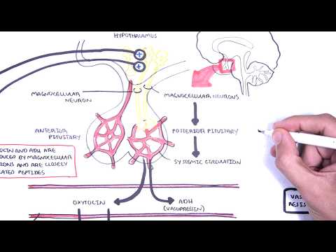
The lecture begins by situating the pituitary gland anatomically beneath the hypothalamus. Dr. Aizaz explains that the pituitary gland, also known as the hypophyseal gland, derives its name from "hypo" meaning "under" and "physis" meaning "growth," highlighting its position as an undergrowth of the hypothalamus. This gland appears as a scrotum-shaped structure at the ventral part of the hypothalamus, though humorously noting it has no relation to the scrotum beyond its shape.
Emphasizing the gland's composition, Dr. Aizaz outlines that the human pituitary gland consists of two main parts:
1. Anterior Pituitary (Anterior Lobe): Also called the adenohypophysis or pars distalis, this is the true glandular component responsible for the synthesis, storage, and release of hormones. Derived from epithelial tissue, it plays a crucial role in endocrine function.
2. Posterior Pituitary (Posterior Lobe): Known as the neurohypophysis or pars nervosa, this is not a true gland but rather an extension of the hypothalamus. It stores and releases hormones produced by the hypothalamus but does not synthesize hormones itself.
The lecture delves into the embryological development of these two parts:
1. Development of the Posterior Pituitary:
The posterior pituitary originates from a downward extension of the hypothalamus. Specifically, an outgrowth from the ventral part of the hypothalamus forms the posterior pituitary. This extension, called the infundibulum, develops into the pituitary stalk and the pars nervosa. Since it is an extension of neural tissue, it is termed the neurohypophysis.
2. Development of the Anterior Pituitary:
The anterior pituitary arises from an upward invagination of the ectodermal tissue from the stomodeum, which is the primitive mouth or oral cavity in the embryo.
This invagination forms a pouch known as Rathke's pouch, named after the scientist who first described it.
Rathke's pouch grows upward toward the neurohypophysis, eventually pinching off from the stomodeum and differentiating into the anterior pituitary. Being derived from ectodermal tissue, the anterior pituitary is an epithelial glandular structure, hence the term adenohypophysis.
Dr. Aizaz provides a detailed explanation of the stomodeum and the oropharyngeal (buccopharyngeal) membrane:
The oropharyngeal membrane is a bilayered structure composed of ectoderm and endoderm with little to no mesoderm between them.
The stomodeum is the ectodermal depression anterior to the oropharyngeal membrane and plays a pivotal role in forming the anterior pituitary.
He clarifies common misconceptions between these two terms, emphasizing that while they are related, they are distinct structures.
Visual 3D animations illustrate the formation and transformation of Rathke's pouch:
Rathke's pouch, initially connected to the stomodeum, eventually loses this connection as it migrates upward.
The cranial part of Rathke's pouch forms the pars tuberalis, which wraps around the pituitary stalk.
The posterior wall contributes to the pars intermedia, a thin layer between the anterior and posterior pituitary, more prominent in other animals than in humans.
The anterior wall becomes the pars distalis, the main hormone-producing region of the anterior pituitary.
Pars Nervosa: Refers to the posterior pituitary/neurohypophysis.
Pars Distalis: Denotes the anterior pituitary/adenohypophysis.
Pars Tuberalis: The collar-like extension of the anterior pituitary that partially encircles the pituitary stalk.
Neurohypophysis vs. Adenohypophysis: Highlighting the neural origin of the posterior pituitary and the glandular origin of the anterior pituitary.
The lecture also touches upon the proximity of the optic chiasma to the pituitary gland, noting that the crossing of optic nerve fibers occurs just ventral to the infundibulum. This anatomical relationship has clinical significance, especially in cases of pituitary tumors affecting vision.
In discussing clinical correlations, Dr. Aizaz mentions:
The potential for remnants of Rathke's pouch to persist along its developmental path. These residual cells can give rise to craniopharyngiomas, benign tumors that may impact surrounding structures due to their location near the optic chiasma and hypothalamus. |
| Category |
Education |
| Tags |
development of pituitary gland | hypophyseal gland | Pituitary gland development | hypophysis | adenohypophysis | neurohypophysis | Rathke's pouch | stomodeum | oropharyngeal membrane | embryology | pars distalis | pars nervosa | pars tuberalis | pars intermedia | craniopharyngioma | optic chiasma | endocrinology | neuroembryology | diencephalon | hypothalamus | prosencephalon |
More Videos