Repair of 50-60% lateral upper eyelid defect w/ free tarsal graft and myocutaneous advancement flap
Hellow guys, Welcome to my website, and you are watching Repair of 50-60% lateral upper eyelid defect w/ free tarsal graft and myocutaneous advancement flap. and this vIdeo is uploaded by Richard C. Allen MD PhD FACS at 2023-05-16T10:16:06-07:00. We are pramote this video only for entertainment and educational perpose only. So, I hop you like our website.
Info About This Video
| Name |
Repair of 50-60% lateral upper eyelid defect w/ free tarsal graft and myocutaneous advancement flap |
| Video Uploader |
Video From Richard C. Allen MD PhD FACS |
| Upload Date |
This Video Uploaded At 16-05-2023 17:16:06 |
| Video Discription |
50% upper eyelid defects can be repaired employing different techniques. Basic principles revolve around repairing the anterior and posterior lamella separately. Often, there is redundant anterior lamella in the upper eyelid that can be used as a myocutaneous flap. Posterior lamellar repair can then be achieved with a free tarsal graft. This way, lid sharing procedures such as a Cutler-Beard can be avoided.
A written transcript of this video is below:
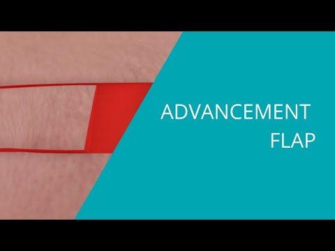
This is Richard Allen at oculosurg.com. This video demonstrates repair of a 50 to 60 percent lateral upper eyelid defect with a free tarsal graft and myocutaneous advancement flap. An anterior lamellar advancement flap is planned with the marking pen. There will be a Burrow’s triangle medially that will be excised. Tissue is excised so that the lid margin section will be horizontal. The Burrow’s triangle is then excised. Dissection is then performed along the length of the eyelid between the orbicularis muscle and the underlying orbital septum to adequately mobilize the flap. This will allow advancement of the anterior lamella. Inspection of the posterior lamella shows that some of the tarsus remains. One could consider advancement of the tarsus; however, the lacrimal gland ductules are adjacent to this area which could result in disruption of the ductules. Therefore, the irregular portion of the tarsus is excised. There remains some lateral tarsus. The posterior lamellar defect is then measured and a free tarsal graft will be harvested from the contralateral upper eyelid. A 15 blade and Westcott scissors are used to harvest the graft. No repair is necessary for the harvest site. The free tarsal graft is then sutured into position with 5-0 Vicryl suture placed partial thickness through the anterior surface of the adjacent tarsus and graft. The upper border of the graft is then sutured to the remaining tarsus with 7-0 Vicryl suture placed in a partial thickness manner. The eyelid margin of the tarsus is then repaired with interrupted 7-0 Vicryl suture placed in a vertical mattress fashion. The anterior lamellar myocutaneous flap is then draped over the posterior lamellar repair. The edges of the skin are sutured together with 5-0 fast absorbing suture. The myocutaneous flap is then sutured to the free tarsal graft with 5-0 Vicryl mattress sutures which are placed partial thickness through the anterior surface of the free tarsal graft. These will be removed at the one week follow up. Additional sutures are placed to realign the skin edges. Inspection of the repair shows the anterior and posterior lamella to be reassociated appropriately. A eye pad will be placed for one day, and the patient will follow up in one week. |
| Category |
Education |
| Tags |
Education Download MP4 | Education Download MP3 | Education Download MP4 360p | Education Download MP4 480p | Education Download MP4 720p | Education Download MP4 1080p |
More Videos